Mechanisms of FMRP
What is the effect of not having any FMRP?
As discussed on the Cytogenetic Analysis page, FMRP is the protein typically produced by FMR1 and has functions discussed here. Fragile X Syndrome manifests in the absence of FMRP (Hagerman et al., 1994).
Many organs and tissues are affected by Fragile X Syndrome because FMRP is typically expressed in all tissues (Singh, Gaur, & Prasad, 2007). In the absence of FMRP, physical phenotypes include a long, narrow face, a slightly enlarged head, a prominent jaw, protruding ears, and macroorchidism (Peier et al., 2000). The main organ system affected by Fragile X Syndrome though is the nervous system because FMRP is primarily expressed in neurons (Singh, Gaur, & Prasad, 2007). The absence of FMRP from the neurons causes the intellectual disability phenotype. It is also primarily expressed in the testes, which contributes to the macroorchidism phenotype (Singh, Gaur, & Prasad, 2007). Behavioral phenotypes include hyperactivity, anxiety, and repetitive behaviors (Gkogkas, 2014). Autism can be an additional phenotype of Fragile X Syndrome, which makes Fragile X Syndrome the most common genetic form of intellectual disability and autism caused by a single gene (Raj, 2021). But what causes all of these phenotypes mechanistically?
Generally speaking, the absence of FMRP from the mutated FMR1 gene causes increased global protein synthesis in affected individuals since FMRP typically acts as a translational repressor (Raj et al., 2021).
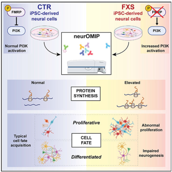
Raj et al. found that the absence of FMRP disrupts phosphoinositide 3-kinase (PI3K) signaling. In Fragile X Syndrome patient-derived neural progenitor cells, p110β expression was increased, which has been found to increase PI3K signaling and global protein synthesis at synapses (Raj et al., 2021). Increased PI3K signaling keeps cells in a proliferative state during neural development, which leads to the observed increased protein synthesis in synapses of the adult brain. The favoring of proliferation over differentiation causes underdeveloped synapses in individuals with Fragile X Syndrome, which causes the intellectual disability associated with the disease. (Raj et al., 2021).
Sharma et al. uncovered a link between overactivated PI3K signaling and overactivated mTOR signaling in Fragile X Syndrome. They found that another activator of PI3K signaling, PIKE-S, is also present in high levels in FMR1 knockout mice. It is a nuclear protein that is an FMRP target. When they added a PI3K inhibitor to mice, it reduced the level of activated mTOR, which confirmed the link between PI3K and mTOR signaling. It makes sense to see a PI3K inhibitor decreasing activated mTOR levels because Sharma et al. found that hippocampal mTOR signaling was increased in FMR1 knockout mice (Sharma et al., 2010). If when PI3K signaling is blocked, which is known to be overactive in Fragile X Syndrome, mTOR activity also decreases, it means they must both be elevated in Fragile X Syndrome. Sharma et al. also observed increased levels of metabotropic glutamate receptor-activated long-term depression (mGluR-LTD), which is a type of synaptic plasticity. mGluR-LTD in synapses requires mammalian target of rapamycin (mTOR), which is a regulator of cell growth, proliferation, and translation (Sharma et al., 2010). If mTOR signaling is overactivated, then mGluR-LTD will also be elevated and proliferation will be favored. In summary, PIKE-S activates PI3K signaling, which activates mTOR signaling, which elevates mGluR-LTD, which causes a preference for cells to proliferate rather than differentiate.
Osterweil et al. connects overactive mGluR5 to overactive ERK1/2 signaling. mGluR5 basally activates ERK1/2, which causes saturation of mRNA translation downstream and leads to elevated protein synthesis in FMR1 knockout mice (Osterweil et al., 2010). This information suggests that ERK1/2 could be another target of Fragile X Syndrome treatment. Osterweil et al. tested an ERK1/2 inhibitor to see if it reversed elevated protein levels associated with Fragile X Syndrome. The results can be found here.
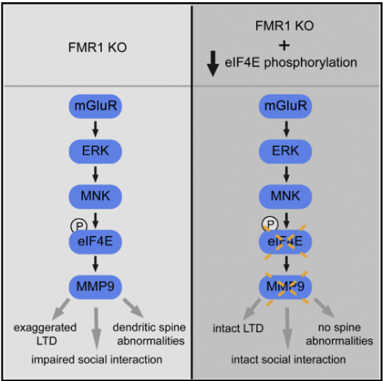
Gkogkas et al. extended upon the work of Sharma et al. and Osterweil et al. that found inhibitory effect of FMRP on translation is regulated by the PI3K/Akt/mTOR and Ras/ERK/MNK pathways. Both pathways stimulate cap-dependent translation by regulating the phosphorylation of transcription factors involved in translation. mTOR promotes the initiation of translation by phosphorylating eukaryotic initiation factor 4E-binding proteins (4E-BPs), which inhibit eIF4E. mTOR also phosphorylates ribosomal protein S6 kinases (S6Ks) to initiate translation. Phosphorylated eIF4E is known to be a regulator of long-lasting forms of synaptic plasticity and memory. It also regulates members of the Matrix Metalloproteinases (MMPs) family, such as Mmp9 mRNA, which affects dendrite morphology, synaptic plasticity, learning, and memory (Gkogkas et al., 2014). FMRP was known to inhibit translation of Mmp9 mRNA in the dendrites, but the mechanism was unknown until Osterweil et al. came along. They found that postmortem brains of individuals with Fragile X Syndrome have increased levels of phosphorylated eIF4E, which regulates MMP-9 and causes high levels of MMP-9 in the brains as well. Overexpression of MMP-9 in mice causes phenotypes associated with Fragile X Syndrome. To connect everything together, Gkogkas et al. found that mGluR activates ERK, which activates MNK, which phosphorylates eIF4E, which regulates MMP-9 production, which is responsible for Fragile X Syndrome phenotypes (Gkogkas et al., 2014).
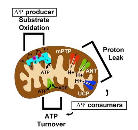
During neural development, neural progenitor cells typically shift from aerobic glycolysis to aerobic respiration during neuronal differentiation, since oxidative phosphorylation is more efficient at producing ATP (Griffiths et al., 2020). Fragile X Syndrome mutants show an increased electron transport chain capacity and higher levels of oxygen consumption, as well as abnormal mitochondrial bioenergetics (Griffiths et al., 2020). When FMRP is not present in cells, there is inefficient oxidative phosphorylation during the peak of synaptogenesis as seen by increased lactate levels and lactate to pyruvate ratios. This is because a deficiency in Coenzyme Q in the forebrain and mitochondrial ubiquinone causes the homeostatic proton leak to be greater than usual, which necessitated FMRP deficient cells to continue relying on aerobic glycolysis to continue producing ATP (Griffiths et al., 2020). The high levels of oxygen consumption observed were a result of cells trying to maintain the mitochondrial membrane potential (Griffiths et al., 2020). The source of the proton leak was mPTP, a nonselective inner mitochondrial membrane mega-channel that regulates synapse maturation and development. When mPTP stays open, it maintains dendritic spines in an immature state, which causes the intellectual disability associated with Fragile X Syndrome (Griffiths et al., 2020). To learn how this proton leak can be fixed, head over to the Treatment Options page!
Works Cited
Gkogkas, C. G.; Khoutorsky, A.; Cao, R.; Jafarnejad, S. M.; Prager-Khoutorsky, M.; Giannakas, N.; Kaminari, A.; Fragkouli, A.; Nader, K.; Price, T. J.; Konicek, B. W.; Graff, J. R.; Tzinia, A. K.; Lacaille, J.-C.; Sonenberg, N. Pharmacogenetic Inhibition of EIF4E-Dependent Mmp9 MRNA Translation Reverses Fragile X Syndrome-like Phenotypes. Cell Reports 2014, 9 (5), 1742–1755. https://doi.org/10.1016/j.celrep.2014.10.064.
Griffiths, K. K.; Wang, A.; Wang, L.; Tracey, M.; Kleiner, G.; Quinzii, C. M.; Sun, L.; Yang, G.; Perez-Zoghbi, J. F.; Licznerski, P.; Yang, M.; Jonas, E. A.; Levy, R. J. Inefficient Thermogenic Mitochondrial Respiration Due to Futile Proton Leak in a Mouse Model of Fragile X Syndrome. The FASEB Journal 2020, 34 (6), 7404–7426. https://doi.org/10.1096/fj.202000283RR.
Hagerman, R. J.; Hull, C. E.; Safanda, J. F.; Carpenter, I.; Staley, L. W.; O’Connor, R. A.; Seydel, C.; Mazzocco, M. M. M.; Snow, K.; Thibodeau, S. N.; Kuhl, D.; Nelson, D. L.; Caskey, C. T.; Taylor, A. K. High Functioning Fragile X Males: Demonstration of an Unmethylated Fully Expanded FMR-1 Mutation Associated with Protein Expression. American Journal of Medical Genetics 1994, 51 (4), 298–308. https://doi.org/10.1002/ajmg.1320510404.
Osterweil, E. K.; Krueger, D. D.; Reinhold, K.; Bear, M. F. Hypersensitivity to MGluR5 and ERK1/2 Leads to Excessive Protein Synthesis in the Hippocampus of a Mouse Model of Fragile X Syndrome. J Neurosci2010, 30 (46), 15616–15627. https://doi.org/10.1523/JNEUROSCI.3888-10.2010.
Peier, A. M.; McIlwain, K. L.; Kenneson, A.; Warren, S. T.; Paylor, R.; Nelson, D. L. (Over)Correction of FMR1 Deficiency with YAC Transgenics: Behavioral and Physical Features. Human Molecular Genetics 2000, 9 (8), 1145–1159. https://doi.org/10.1093/hmg/9.8.1145.
Raj, N.; McEachin, Z. T.; Harousseau, W.; Zhou, Y.; Zhang, F.; Merritt-Garza, M. E.; Taliaferro, J. M.; Kalinowska, M.; Marro, S. G.; Hales, C. M.; Berry-Kravis, E.; Wolf-Ochoa, M. W.; Martinez-Cerdeño, V.; Wernig, M.; Chen, L.; Klann, E.; Warren, S. T.; Jin, P.; Wen, Z.; Bassell, G. J. Cell-Type-Specific Profiling of Human Cellular Models of Fragile X Syndrome Reveal PI3K-Dependent Defects in Translation and Neurogenesis. Cell Rep 2021, 35 (2), 108991. https://doi.org/10.1016/j.celrep.2021.108991.
Sharma, A.; Hoeffer, C. A.; Takayasu, Y.; Miyawaki, T.; McBride, S. M.; Klann, E.; Zukin, R. S. Dysregulation of MTOR Signaling in Fragile X Syndrome. J. Neurosci. 2010, 30 (2), 694–702. https://doi.org/10.1523/JNEUROSCI.3696-09.2010.
Singh, K.; Gaur, P.; Prasad, S. Fragile x Mental Retardation (Fmr-1) Gene Expression Is down Regulated in Brain of Mice during Aging. Mol Biol Rep 2007, 34 (3), 173–181. https://doi.org/10.1007/s11033-006-9032-8.

Very interesting topic!!
Thank you! I’m glad that you enjoyed it!