Reflection Blog 3: An Outline
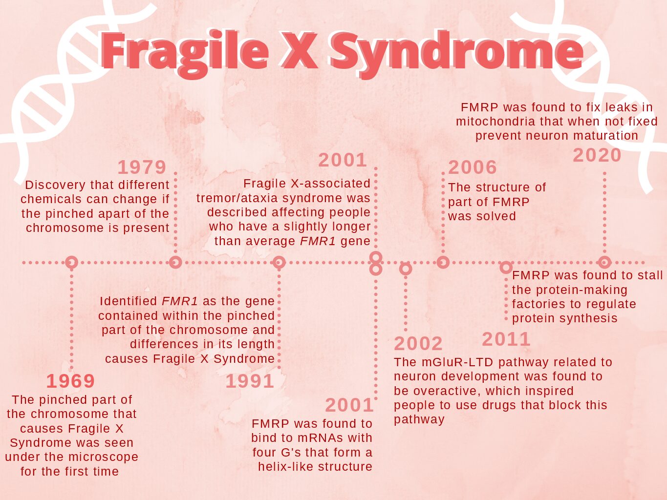
Disease Page 1: Cytogenetic Analysis This page will address the question, “What does Fragile X Syndrome look like under the microscope?” I want to start with a brief history to provide context for why people began to perform cytogenetic analysis that led to the discovery of the fragile site, which will come from the history section of Defining the Disease. The cytogenetic analysis of the X chromosome started because of the observation that most intellectually disabled individuals were men. I will then talk about the first paper exploring Fragile X Syndrome, which was the landmark paper “A marker X chromosome” by Lubs et al. in 1969. They were the first people to observe the fragile site. The history section of Defining the Disease will be used to summarize their work. A landmark paper from 1979 titled, “Heritable fragile sites in human chromosomes I. Factors affecting expression in lymphocyte culture” by Sutherland found that the appearance of the fragile site differs in different conditions. That paper’s findings will be summarized here.
“Instability of a 550-Base Pair DNA Segment and Abnormal Methylation in Fragile X Syndrome” by Oberlé et al. and “Identification of a gene (FMR-1) containing a CGG repeat coincident with a breakpoint cluster region exhibiting length variation in fragile X syndrome” by Verkerk et al. were two other landmark papers that identified the gene behind Fragile X Syndrome. These papers will be discussed as they identified the gene at the fragile site.
I will then talk about what causes the fragile site biochemically, which was outlined in the disease state cellular, biomolecular and atomic sections of Defining the Disease.
At this point, there will be a link to the FMRP protein page where I will discuss the discovery of the gene affected by the fragile site that produces FMRP to connect those two pages together.
Figures included will be images of the fragile site under a microscope and perhaps a cartoon illustration of the hydrogen bonds that form to make the G-quadruplex in the CGG repeats that cause the fragile site.
Disease Page 2: FMRP Protein The star of the show is the protein behind Fragile X Syndrome – FMRP. I will talk about one of the first papers to try and identify the protein involved in Fragile X Syndrome, which was “Fragile X syndrome: search for phenotypic manifestations at loci for hypoxanthine phosphoribosyltransferase and glucose-6-phosphate dehydrogenase” by Mareni and Migeon. However, they were unsuccessful. I will then talk about Feng et al.’s findings from “Fragile X Mental Retardation Protein: Nucleocytoplasmic Shuttling and Association with Somatodendritic Ribosomes” that identified where FMRP is located. I will also use my summaries from the healthy state cellular and biomolecular sections of Defining the Disease to further talk about where FMRP is located.
I will then move onto talking about the structure of FMRP, which I initially talked about in the last question of Defining the Disease. A paper by Bardoni, Schenck, and Louis Mandel titled, “A Novel RNA-binding Nuclear Protein That Interacts With the Fragile X Mental Retardation (FMR1) Protein” talks about some protein-protein interactions that were also highlighted in the last question of Defining the Disease, so I will briefly talk about protein-protein interactions here. There is a paper by Zalfa et al. titled “The Fragile X Syndrome Protein FMRP Associates with BC1 RNA and Regulates the Translation of Specific mRNAs at synapses that I will summarize because it talks about how FMRP may be directed to the mRNAs it targets. I will then segue and provide a link to the mechanism page for people to dive more into the biochemistry of it.
Figures on this page will include pictures of the cells that contain FMRP as well as the structural images I annotated in Chimera for question four of Defining the Disease.
Disease Page 3: Mechanisms of FMRP This page will outline how FMRP works. Most of the information will come from the subcellular and atomic disease state and healthy state sections of Defining the Disease. “Hypersensitivity to mGluR5 and ERK1/2 Leads to Excessive Protein Synthesis in the Hippocampus of a Mouse Model of Fragile X Syndrome” by Osterweil et al. talks about FMRP’s relationship to the mGluR5 and ERK1/2 signaling pathways. One of the landmark papers that will be highlighted in this section is “FMRP stalls ribosomal translocation on mRNAs linked to synaptic function and autism” by Darnell et al. It discusses how FMRP acts as a translational repressor. “Pharmacogenetic Inhibition of eIF4E-Dependent Mmp9 mRNA Translation Reverses Fragile X Syndrome-like Phenotypes” by Gkogkas et al. talks about FMRP’s connection to the Ras/ERK/Mnk pathway. Another landmark paper that talks about FMRP’s mechanism is “ATP synthase c-subunit leak causes aberrant cellular metabolism in Fragile X syndrome” by Licznerski et al., which discusses how the absence of FMRP causes a normal physiological proton leak to become exaggerated and pathologic.
Figures will include graphics of the pathways that FMRP affects and where in the pathway it acts.
Healthy Page: The healthy page will talk about the normal function of FMRP. The majority of this information will come from the healthy state section of Defining the Disease. The main result of the absence of FMRP is the prevention of the formation of mature dendrites, so I will talk about how dendrites normally form and the importance of their formation in learning and memory. I will also reiterate where FMRP is typically found. The bulk of the page will be describing the normal functioning of the pathways that FMRP affects. This includes the normal metabolic switch from aerobic glycolysis to oxidative phosphorylation during neuronal differentiation. This also includes talking about the normal functioning of the proton leak in the electron transport chain.
Figures will include images of mature dendrites, a graphic that depicts the shift to oxidative phosphorylation, and a graphic that shows the proton leak in the electron transport chain.
Works Cited
Lubs, H. A. A marker X chromosome. Am J Hum Genet 21, 231–244 (1969).