In the Absence of Fragile X Syndrome
What is the “normal” functioning of people without Fragile X Syndrome?
The main organ system affected by Fragile X Syndrome is the nervous system. In a healthy state, the nervous system can effectively send messages between neurons to facilitate successful learning and consolidate memories (Arikkath, 2012). Many physical phenotypes associated with Fragile X Syndrome pertain to body parts being proportioned differently (Peier et al., 2000). In a healthy state, the face is wider, the head and testes are smaller, and the ears and jaw protrude less than in the disease state.
In the healthy state, a secondary constriction site, called a fragile site, is not present in any cells on chromosome Xq27.3 (Lubs et al.,1969). To see what the chromosome looks like with the fragile site, click here. The 5’ untranslated region of the FMR1 gene contains 5-50 unmethylated CGG repeats, which does not form any three-dimensional structure that inhibits DNA replication or transcription. This allows the FMR1 gene to be transcribed and produce fragile X mental retardation protein (FMRP) at normal levels.
FMRP is found in all cells, but primarily in the cytoplasm, nucleoplasm, nuclear pores, and dendrites of neurons (Feng et al., 1997). It is synthesized in the cytoplasm, then moves to the nucleus where it associates with other proteins, RNAs, and ribosomes. It then goes back out into the cytoplasm where it is detected as part of a large ribosome-containing ribonucleoprotein particle and transported along the dendrites to the neural synapses (Ramos et al., 2006). The protein was found to primarily co-fractionate with intraneuronal polysomes (Feng et al., 1997). FMRP is also found in astrocytes, microglia, and oligodendrocytes early in life (Griffiths et al., 2020). Specifically, Griffiths et al. observed a peak in steady-state FMRP levels during the tenth day of life in mice. Additionally, FMRP is found in testes (Singh, Gaur, & Prasad, 2007).
FMRP is found in different areas of the cell because it facilitates the shuttling of mRNAs between the nucleus and cytoplasm, which is known because it has a nuclear export signal as well as a nuclear localization signal (Ramos et al., 2006; Singh, Gaur, & Prasad, 2007). The N-terminus of FMRP contains the nucleolar localization signal, which was determined through a crystal structure solved by Ramos et al. Residues T125 and F126 (highlighted in orange in Figure 1) in the ⍺ helix were found to be critical in determining the stability of the NDF fold and therefore the localization of FMRP. They hypothesize that the NDF may allosterically regulate the functions of FMRP and what it will bind depending on its localization since as mentioned above it can exist in the cytoplasm or the nucleus (Ramos et al., 2006).
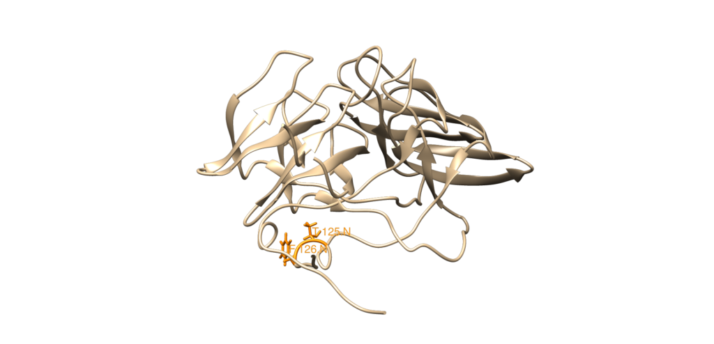
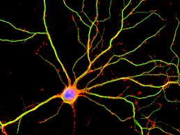
Ramos et al. found that the N-terminal domain of FMRP (NDF), which makes up the first 134 residues of the N-terminus, contains two repeats of a Tudor domain and a short, C-terminal ⍺ helix, which interact together to stabilize the composite NDF fold in a compact domain. The Tudor repeats were found to be flexible and fold into a bent, four-stranded, antiparallel 𝛽 sheet with another 𝛽 sheet closing the cavity. The Tudor repeats formed hydrophobic pockets that Ramos et al. believe play a role in protein-protein interactions. FMRP had already been known to interact with 82-FIP and NUFIP in the nucleus. One of the Tudor repeats of the NDF was found to interact with methylated lysine and 82-FIP. FMRP also plays a role in neural development to differentiate neuronal cells and create mature dendrites (Raj et al., 2021). Dendrites are the part of neurons that contain receptors and channels to receive chemical information from the synapses sent by other neurons (Arikkath, 2012). In other words, they receive communication from other neurons. Dendrites arise from the soma of neurons during neural development (Alberts et al., 2002). If the dendrites do not fully emerge from the neuron and form synapses with other neurons, then neurons cannot communicate with each other, which has adverse effects on memory and learning (Arikkath, 2012).
FMRP also typically acts as a translational repressor, so in the healthy state, translation and protein levels are regulated and neural cell differentiation is favored over proliferation (Raj et al., 2021). For example, PIKE-S mRNA, which activates PI3K signaling, is a target of FMRP (Sharma et al., 2010). FMRP binds to the mRNA to keep PI3K signaling controlled and decrease proliferation (Sharma et al., 2010). FMRP binds to mRNAs that code for synaptic plasticity-related proteins primarily targeted by a G quartet structure and represses translation of those mRNAs by directly stalling ribosomal translation during elongation in a complex of FMRP, target mRNAs, and stalled ribosomes (Darnell et al., 2011). Some of these proteins found to be targets of FMRP are the product encoded by the NPTXR mouse gene, SAPAP4, and DAP-1 (Brown et al., 2001; Darnell et al., 2011). NPTXR codes for neuronal pentraxin receptor, which is a facilitator of dendrite growth and synapse formation (Atluri et al., 2013). SAPAP4 and DAP-1 maintain postsynaptic density and play a role in neuronal cell signaling (Brown et al., 2001).
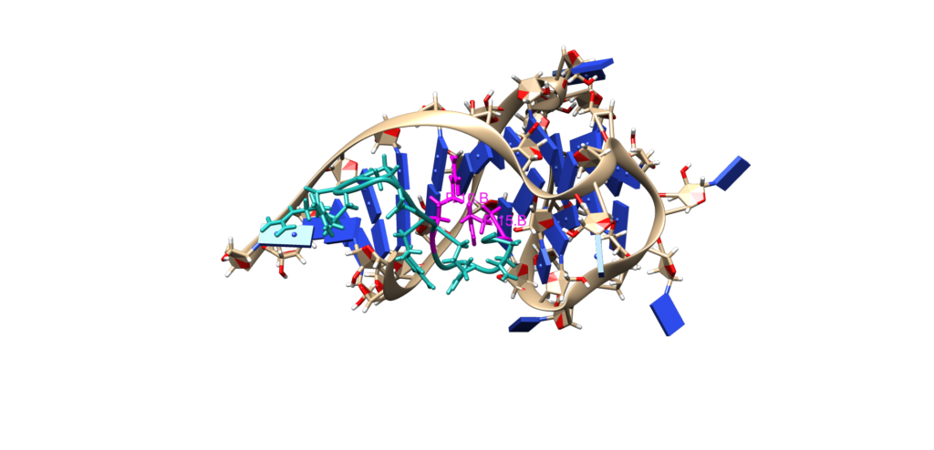
Phan et al. solved the crystal structure of an FMRP arginine-glycine-rich RGG peptide (colored in teal in Figure 3) with guanine-rich sc1 RNA (colored tan, red, and blue in Figure 3) to understand how FMRP targets and binds to its RNA targets (Phan et al., 2011). The small FMRP peptide was used and not the whole protein because only the small arginine-glycine rich RGG box is necessary to bind guanine-rich RNAs. Both the peptide and RNA are unstructured in the free state, but become structured in the bound state. The RNA formed a G-quadruplex when bound to the FMRP peptide and the complex formed a duplex-quadruplex junction (Phan et al., 2011). R10 and R15 (colored magenta in Figure 3) are responsible for recognizing the duplex-quadruplex junction and situating the RNA into the binding pocket of the protein. There is evidence that this complex can be regulated because methylation of R10 and/or R15 results in a decrease in binding RNA (Phan et al., 2011).
FMRP is also known to bind the mRNA of all the enzymes involved in Coenzyme Q synthesis, so it may influence CoQ production, which is essential for the electron transport chain (Griffiths et al., 2020). Before neural progenitor cells are mature, they use aerobic glycolysis to produce ATP. During neuronal differentiation, a metabolic switch occurs to primarily use aerobic respiration because oxidative phosphorylation is more efficient at producing ATP (Griffiths et al., 2020). The switch is also important in neuron maturation, synaptogenesis, and synaptic protein translation (Griffiths et al., 2020). mPTP, a nonselective inner mitochondrial membrane mega-channel, typically opens transiently during nervous system development to generate mitochondrial superoxide that suppresses neural progenitor cell proliferation and promotes differentiation and the switch from aerobic glycolysis to aerobic respiration. Thus, it typically acts as a regulator of synapse maturation and development (Griffiths et al., 2020).
A slight proton leak is a normal physiological process that causes an incomplete coupling between generating ATP and moving electrons through the electron transport chain in oxidative phosphorylation. This incomplete coupling allows some cells to generate heat, maintain carbon flux, and alter nutrient response in different conditions. It also works to regulate mitochondrial membrane potential to prevent oxidative stress. In FMR1 knockout mice, this proton leak is exaggerated to the point that it becomes pathologic (Griffiths et al., 2020).
To learn how these pathways are affected in Fragile X Syndrome, head to the mechanisms page here.
Works Cited
Alberts, B.; Johnson, A.; Lewis, J.; Raff, M.; Roberts, K.; Walter, P. Neural Development. Molecular Biology of the Cell. 4th edition 2002.
Arikkath, J. Molecular Mechanisms of Dendrite Morphogenesis. Frontiers in Cellular Neuroscience 2012, 6.
Atluri, V. S. R.; Kanthikeel, S. P.; Reddy, P. V. B.; Yndart, A.; Nair, M. P. N. Human Synaptic Plasticity Gene Expression Profile and Dendritic Spine Density Changes in HIV-Infected Human CNS Cells: Role in HIV-Associated Neurocognitive Disorders (HAND). PLoS One 2013, 8 (4), e61399. https://doi.org/10.1371/journal.pone.0061399.
Brown, V.; Jin, P.; Ceman, S.; Darnell, J. C.; O’Donnell, W. T.; Tenenbaum, S. A.; Jin, X.; Feng, Y.; Wilkinson, K. D.; Keene, J. D.; Darnell, R. B.; Warren, S. T. Microarray Identification of FMRP-Associated Brain MRNAs and Altered MRNA Translational Profiles in Fragile X Syndrome. Cell 2001, 107 (4), 477–487. https://doi.org/10.1016/S0092-8674(01)00568-2.
Darnell, J. C.; Van Driesche, S. J.; Zhang, C.; Hung, K. Y. S.; Mele, A.; Fraser, C. E.; Stone, E. F.; Chen, C.; Fak, J. J.; Chi, S. W.; Licatalosi, D. D.; Richter, J. D.; Darnell, R. B. FMRP Stalls Ribosomal Translocation on MRNAs Linked to Synaptic Function and Autism. Cell 2011, 146 (2), 247–261. https://doi.org/10.1016/j.cell.2011.06.013.
Feng, Y.; Gutekunst, C.-A.; Eberhart, D. E.; Yi, H.; Warren, S. T.; Hersch, S. M. Fragile X Mental Retardation Protein: Nucleocytoplasmic Shuttling and Association with Somatodendritic Ribosomes. J. Neurosci. 1997, 17 (5), 1539–1547. https://doi.org/10.1523/JNEUROSCI.17-05-01539.1997.
Griffiths, K. K.; Wang, A.; Wang, L.; Tracey, M.; Kleiner, G.; Quinzii, C. M.; Sun, L.; Yang, G.; Perez-Zoghbi, J. F.; Licznerski, P.; Yang, M.; Jonas, E. A.; Levy, R. J. Inefficient Thermogenic Mitochondrial Respiration Due to Futile Proton Leak in a Mouse Model of Fragile X Syndrome. The FASEB Journal 2020, 34 (6), 7404–7426. https://doi.org/10.1096/fj.202000283RR.
Lubs, H. A. A Marker X Chromosome. Am J Hum Genet 1969, 21 (3), 231–244.
Peier, A. M.; McIlwain, K. L.; Kenneson, A.; Warren, S. T.; Paylor, R.; Nelson, D. L. (Over)Correction of FMR1 Deficiency with YAC Transgenics: Behavioral and Physical Features. Human Molecular Genetics 2000, 9 (8), 1145–1159. https://doi.org/10.1093/hmg/9.8.1145.
Phan, A. T.; Kuryavyi, V.; Darnell, J. C.; Serganov, A.; Majumdar, A.; Ilin, S.; Raslin, T.; Polonskaia, A.; Cynthia Chen; Clain, D.; Darnell, R. B.; Patel, D. J. Structure-Function Studies of FMRP RGG Peptide Recognition of an RNA Duplex-Quadruplex Junction. Nature Structural & Molecular Biology 2011, 18 (7), 796–804. https://doi.org/10.1038/nsmb.2064.
Raj, N.; McEachin, Z. T.; Harousseau, W.; Zhou, Y.; Zhang, F.; Merritt-Garza, M. E.; Taliaferro, J. M.; Kalinowska, M.; Marro, S. G.; Hales, C. M.; Berry-Kravis, E.; Wolf-Ochoa, M. W.; Martinez-Cerdeño, V.; Wernig, M.; Chen, L.; Klann, E.; Warren, S. T.; Jin, P.; Wen, Z.; Bassell, G. J. Cell-Type-Specific Profiling of Human Cellular Models of Fragile X Syndrome Reveal PI3K-Dependent Defects in Translation and Neurogenesis. Cell Rep 2021, 35 (2), 108991. https://doi.org/10.1016/j.celrep.2021.108991.
Ramos, A.; Hollingworth, D.; Adinolfi, S.; Castets, M.; Kelly, G.; Frenkiel, T. A.; Bardoni, B.; Pastore, A. The Structure of the N-Terminal Domain of the Fragile X Mental Retardation Protein: A Platform for Protein-Protein Interaction. Structure 2006, 14 (1), 21–31. https://doi.org/10.1016/j.str.2005.09.018.
Sharma, A.; Hoeffer, C. A.; Takayasu, Y.; Miyawaki, T.; McBride, S. M.; Klann, E.; Zukin, R. S. Dysregulation of MTOR Signaling in Fragile X Syndrome. J. Neurosci. 2010, 30 (2), 694–702. https://doi.org/10.1523/JNEUROSCI.3696-09.2010.
Singh, K.; Gaur, P.; Prasad, S. Fragile x Mental Retardation (Fmr-1) Gene Expression Is down Regulated in Brain of Mice during Aging. Mol Biol Rep 2007, 34 (3), 173–181. https://doi.org/10.1007/s11033-006-9032-8.

You mention on this page that FMRP is known to bind to the mRNA that interacts with CoQ. Is there any data that this interaction can lead to an increase in radical oxygen production (increase in oxidative stress)?
Hi, Zach! Thanks for this great question! The paper linked here was the first paper to detect higher than normal levels of reactive oxygen species in FMR1 knockout mice and thus increased oxidative stress.